 In modern medicine, medical imaging has undergone major advancements. Today, this ability to achieve information about the human body has many useful clinical applications. Over the years, different sorts of medical imaging have been developed, each with their own advantages and disadvantages.
X-ray based methods of medical imaging include conventional X-ray, computed tomography (CT) and mammography. To enhance the X-ray image, contrast agents can be used for example for angiography examinations.
Molecular imaging is used in nuclear medicine and uses a variety of methods to visualize biological processes taking place in the cells of organisms. Small amounts of radioactive markers, called radiopharmaceuticals, are used for molecular imaging.
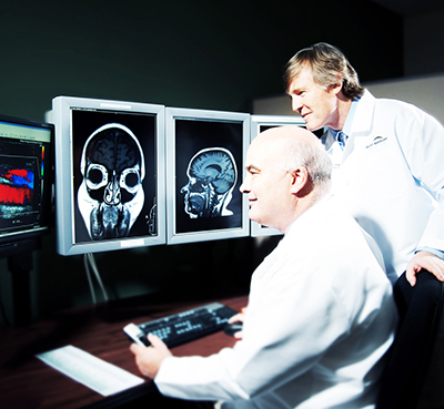
Other types of medical imaging are magnetic resonance imaging (MRI) and ultrasound imaging. Unlike conventional X-ray, CT and Molecular Imaging, MRI and ultrasound operate without ionizing radiation. MRI uses strong magnetic fields, which produce no known irreversible biological effects in humans.
Diagnostic ultrasound systems use high-frequency sound waves to produce images of soft tissue and internal body organs.
When imaging with X-rays, an X-ray beam produced by a so-called X-ray tube passes through the body. On it’s way through the body, parts of the energy of the X-ray beam are absorbed. This process is described as attenuation of the X-ray beam. On the opposite side of the body, detectors or a film capture the attenuated X-rays, resulting in a clinical image. In conventional radiography, one 2D image is produced. In Computed Tomography, the tube and the detector are both rotating around the body during the examination so that multiple images can be acquired, resulting in a 3D visualization.
The most common methods of X-ray in medical imaging are X-ray radiography, computed tomography (CT), mammography, angiography and fluoroscopy.
Different organs and tissues have a different sensitivity to radiation. This is why the actual risk to the body from X-ray procedures varies depending on the part of the body being X-rayed. “Effective dose” is a parameter of the dose absorbed by the entire body that takes account of these differing sensitivities.
Doctors and manufacturers are well aware of the risks and do everything possible to minimize radiation dose. Guided by technical standards that are set and continually updated by national and international radiology protection councils, they take special care during X-ray examinations to use the lowest radiation dose possible while producing the images for. Advanced X-ray systems contain special features that help reduce the radiation dose. For example there are technologies developed to ensure that those parts of a patient’s body not being imaged receive no or only minimal radiation exposure.